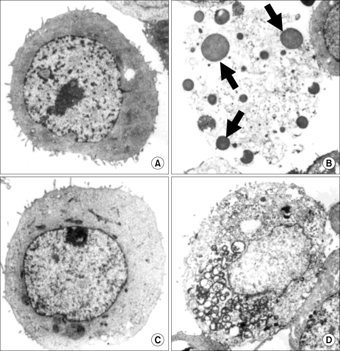
Fig. 4.
Electron microphotographs of MKN-45 and MKN-28 cells after treatment with urushiol. After treatment with urushiol morphological changes of MKN-45 and MKN-28 cells were observed by electron microscopy. Untreated control of MKN-45 cells (A); 15 μg/ml urushiol-treated MKN-45 cells (B); untreated control of MKN-28 cells (C); 20 μg/ml urushiol-treated MKN-28 cells (D). Multiple nuclear fragments (arrows) were observed on urushiol-treated MKN-45 cells (B).

