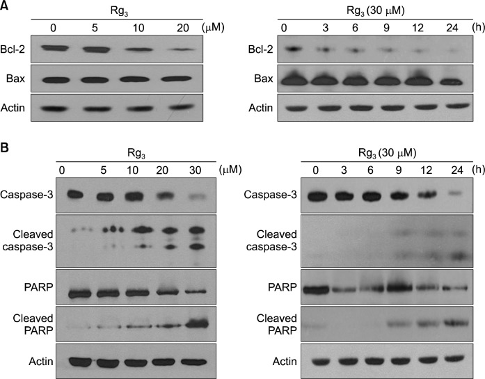
Fig. 3.
Effects of Rg3 on the expression of the apoptotic markers. MDA-MB-231 cells were treated with Rg3 (5, 10, 20, 30 μM) for 24 h (left panel). MDA-MB-231 cells were treated with 30 μM of Rg3 for the time indicated (right panel). The levels of Bcl-2 and Bax (A) as well as caspase-3, cleaved caspase-3, PARP, and cleaved PARP (B) were determined by Western blot analysis.

