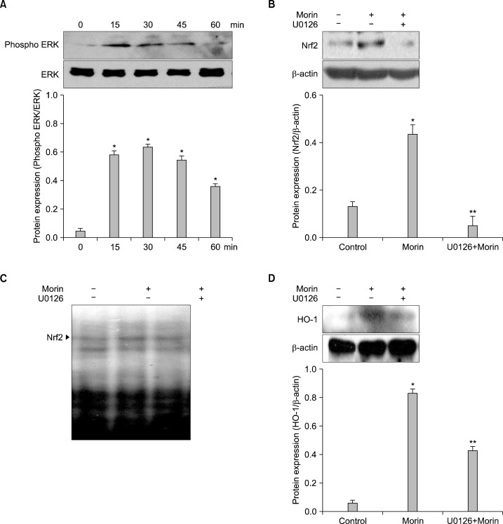
Fig. 4.
Morin up-regulates HO-1 via phosphorylation of ERK. Cells were treated with morin for various times. (A) Western blot were performed to detect expression of ERK. *Significantly different from control cells (P<0.05). Cells were pre-incubated with inhibitor-U0126 (2.5 μM) for 1 h and the protein was analyzed by (B) western blot and (C) DNA binding activity was detected by EMSA. *Significantly different from control cells (P<0.05). **Significantly different from morin-treated cells (P<0.05). (D) Cells were pre-incubated with U0126 for 1 h, and after incubation of 8 h, the HO-1 expression was analyzed by western blot. *Significantly different from control cells (P<0.05). **Significantly different from morin-treated cells (P<0.05).

