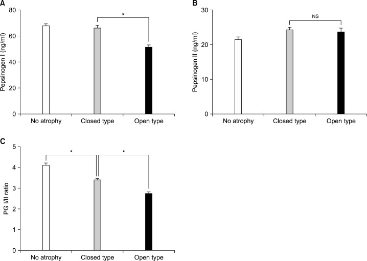
Fig. 2.
The serum pepsinogen (PG) levels depending on the distribution of atrophic gastritis by endoscopy. The level of serum PG I was significantly lower in open-type atrophic gastritis, closed-type atrophic gastritis (A). In contrast, serum PG II levels did not show any significant differences among groups (B). Serum PG I/II ratio decreased significantly as endoscopic gastric mucosal atrophy progressed (C). Data are presented as mean±S.E. *P<0.001; NS, not significant.

