Figure 1.
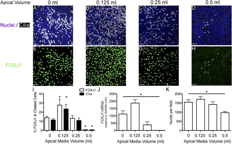
Forkhead box J1 (FOXJ1) expression and ciliated cell differentiation are reduced by submersion in an apical volume–dependent manner. (A–H) Representative extended focus confocal immunofluorescent images of NHBE cells cultured for 21 days with apical media volumes of 0 ml (A and E), 0.125 ml (B and F), 0.25 ml (C and G), and 0.5 ml (D and H) stained for nuclei (Hoechst, blue) and cilia (acetylated α-tubulin, white; A–D) or FOXJ1 (green; E–H). (I) Percent FOXJ1-positive (FOXJ1+; white bars) and ciliated (black bars) cells at the apical media volumes, indicated on the x axis, showing a significant increase at 0.125 ml and a significant decrease at 0.5 ml. (J) FOXJ1 messenger RNA (mRNA) levels after 21 days of differentiation at the apical media volumes, indicated on the x axis, normalized to glyceraldehyde phosphate dehydrogenase (GAPDH) mRNA, showing that the amount of FOXJ1 mRNA increases at 0.125 ml and decreases significantly at higher apical volumes, which is similar to changes in FOXJ1+ cells. (K) Quantification of nuclear density after 21 days submerged with different volumes of media showing a significant reduction in cells in 0.5 ml. Scale bar, 50 μm. Data shown are means (± SEM). One-way ANOVA (*P < 0.05; n = 3).

