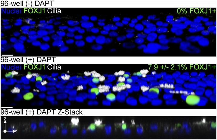
Figure 6.
DAPT enables FOXJ1 expression and ciliated cell differentiation of NHBE cells in submerged culture on plastic in 96-well plates. Representative three-dimensional (3D) opacity renderings of 40× confocal immunofluorescent images of NHBE cells cultured in wells of a 96-well plate submerged for 21 days (top) or submerged with 10 μM DAPT (middle); a Z-stack of submerged conditions with 10 μM DAPT is also shown (bottom). Cells were stained for cilia (acetylated α-tubulin, white), FOXJ1 (green), and nuclei (Hoechst, blue). The percent FOXJ1+ cells are indicated in the upper right corner of the upper two panels. The 3D images were cropped and rotated along the z axis to view cells from above the apical surface. Scale bar, 20 μm (n = 3).

