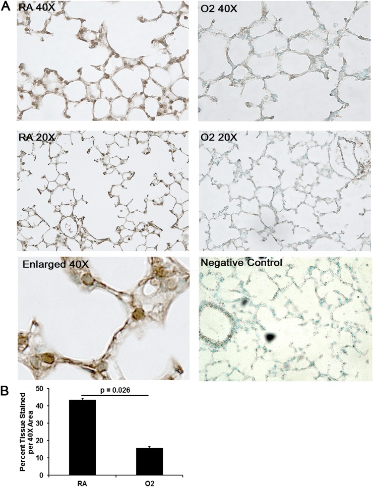
Figure 2.
Immunohistochemistry (IHC) of extracellular superoxide dismutase (SOD)3 in RA- versus O2-exposed mice. (A) Brown 3,3'-diaminobenzidine staining denotes positive staining of SOD3 on 20× and 40× magnification. Representative images of RA-exposed mice demonstrate continuous staining of SOD3 along the extracellular matrix, whereas O2-exposed (5 d) mice demonstrate fragmented and decreased deposition of SOD3 along the extracellular matrix. (B) Quantitative IHC confirms reduced SOD3 deposition in O2-exposed lungs (n = 3 per group) (P = 0.026).

