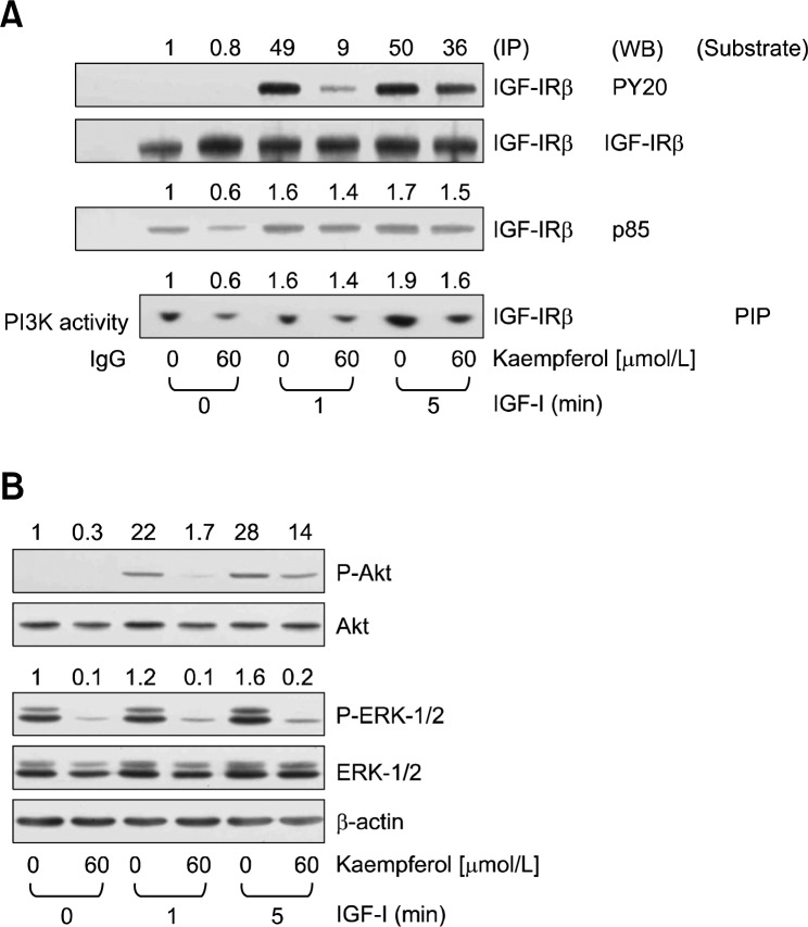
Figure 4.
Effect of kaempferol on the insulin-like growth factor-I receptor (IGF-IR) signaling pathway. HT-29 cells were treated with 0 or 60 μmol/L kaempferol for 2 hours and lysed after 0, 1, or 5 minutes of stimulation with 10 nmol/L insulin-like growth factor-I (IGF-I). (A) Total cell lysates were incubated with anti-IGF-IRβ antibody and the immune complexes were precipitated with protein A-Sepharose. The immunoprecipitated proteins were subjected to Western blot analysis with the relevant antibodies. The immunoprecipitated proteins were incubated with phosphatidylinositol and [γ-32P]ATP to detect phosphatidylinositol 3-kinase (PI3K) activity. The resulting 32P-labelled phosphatidylinositol 3-phosphate (PIP) was separated by thin-layer chromatography and visualized by autoradiography. (B) Total cell lysates were subjected to Western blot analysis with their relevant antibodies. ERK, extracellular regulated kinase; IP, immunoprecipitation; WB, western blot analysis.

