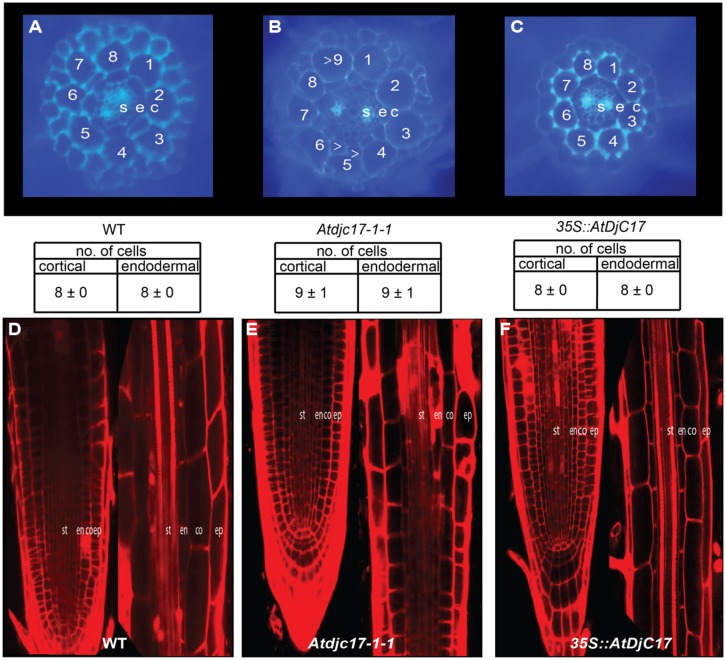
FIGURE 5.
Examination of cell division in WT, Atdjc171-1 and 35S::AtDjC17 roots: agarose embedded hand sections stained with calcofluor-white stained and confocal microscopic images of propidium iodide stained WT, Atdjc17-1-1 and 35S::AtDjC17 mutant roots. (A) WT section. (B) Atdjc17-1-1 section showing additional cell division in the cortical and endodermal layers. Note that the divisional pattern seems anticlinal in nature. Arrow heads indicate ectopic divisions. (C) 35S::AtDjC17 section with no evidence of altered cortical and endodermal cell numbers. (D) 7-day old post-germination WT root tip and zone above the meristematic tip in WT root. (E) Atdjc17-1-1 root tip with associated zone above the meristematic tip.(F) 35S::AtDjC17 root tip and elongation zone. st, stele; en, endodermis; co, cortex; ep, epidermis.

