
Figure 8.
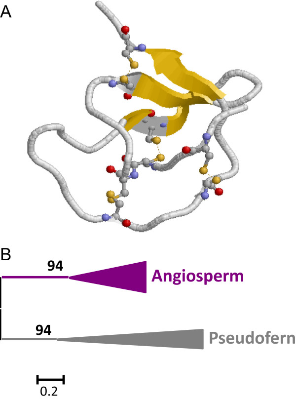
Features of I20 Pin-II peptidase inhibitors. (A) Three-dimensional structure of a typical I20 inhibitor (4SGB). Cysteines are highlighted as balls and sticks and coloured in CPK. Yellow, β-sheets. (B) Schematic PhyML phylogenetic tree using the selected Pin-II sequences from the different plant species. Coloured triangles show clade-specific gene proliferations.