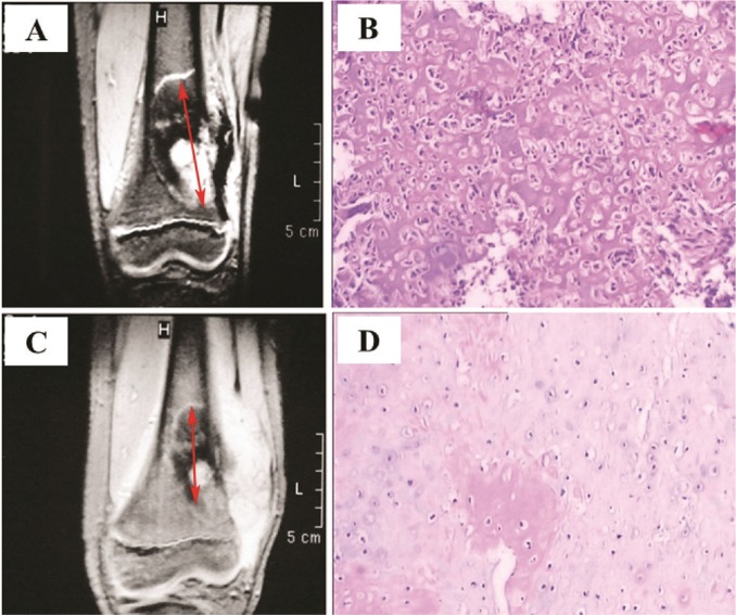
FIGURE 3.
Osteosarcoma arising from the left distal femur in a 15-year-old boy. (A) Magnetic resonance image obtained before neoadjuvant chemotherapy. Arrows show longest diameter of lesion. (B) Tumour cells from the lesion. Hematoxylin and eosin, 40× original magnification. (C) Magnetic resonance image of the same tumour after chemotherapy. Calcification is extensive, and the lesion (arrow) is significantly smaller (≥30% reduction), indicating a good response to therapy. (D) Tumour cells show significant degeneration and necrosis. Hematoxylin and eosin, 40× original magnification. The patient was evaluated as having a partial response by the Response Evaluation Criteria in Solid Tumors (version 1.1).

