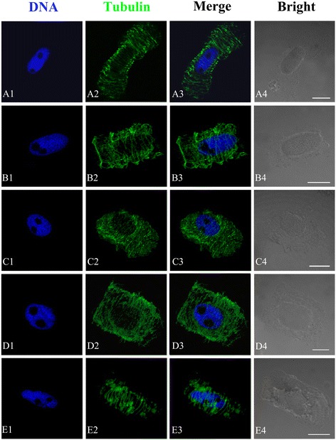
Figure 4.

Effects of different concentrations of Al on the organization of microtubule cytoskeleton in root tip cells of P. massoniana . DNA staining with DAPI (A1–E1, blue), tubulin immunolabeling (A2–E2, green), merged images (A3–E3) and bright field images (A4–E4) in the same single optical section obtained with the confocal laser scanning microscope. Bar = 10 μm for all figures. A. Interphase cells. The nucleus is surrounded by numerous cortical MTs that are orientated transversely to the long cell axis. B. Showing aberrant cortical MTs with slightly skewed wavy (10−5 M Al, 24 h). C–D. Showing numerous interconnected and short MT fragments distributed randomly at the cell periphery (10−4 M Al, 48 h). E. Showing MT stickiness (10−3 M Al, 48 h).
