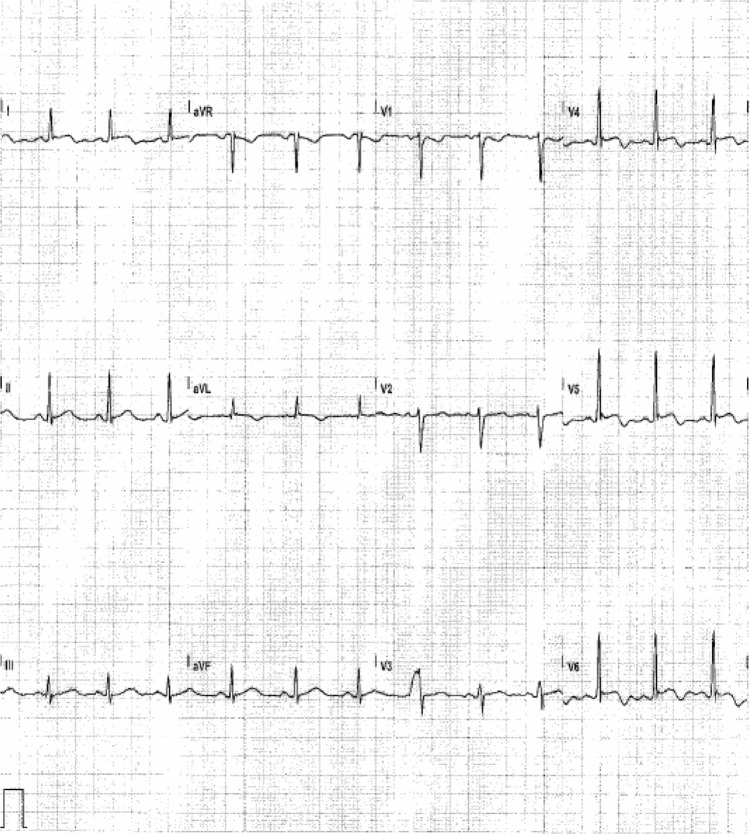
Figure 1.
First cardiac event.
Notes: Traces from the 12-lead ECG (25 mm/s, 10 mm/mV) showed transient abnormalities. Peaked T wave and ST segment elevation were associated with thoracoabdominal discomfort. I, II, and III = limb leads; V1 to V6 = precordial leads.
Abbreviations: aVF, lead augmented vector foot; aVL, lead augmented vector left; aVR, lead augmented vector right; ECG, electrocardiogram.

