
Figure 6.
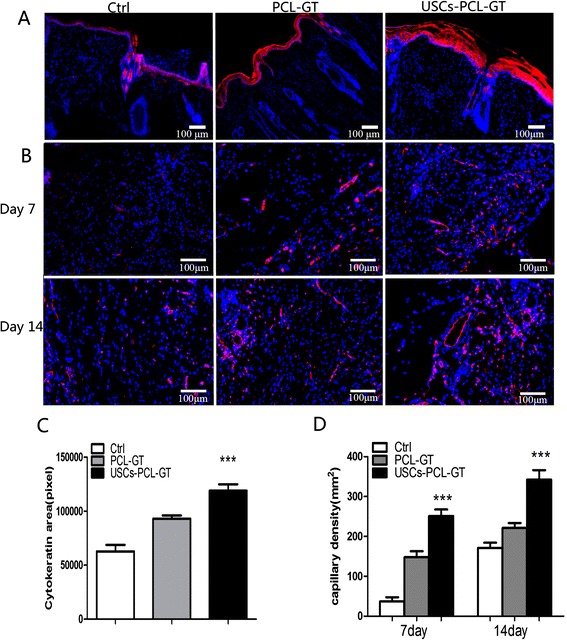
Tissue sections were immunostained with anti-pan-cytokeratin (A) and anti-CD31 antibodies (B). Nuclei (blue) were stained with DAPI. (C) Significantly greater cytokeratin area was observed in the USCs-PCL/GT group (***P < 0.01 compared to the control group, *P < 0.05 compared to the PCL/GT group). (D) Significantly increased microvessel density was observed in the USCs-PCL/GT group (***P < 0.01 compared to the control and PCL/GT groups). Scale bar = 100 μm.