
Figure 7.
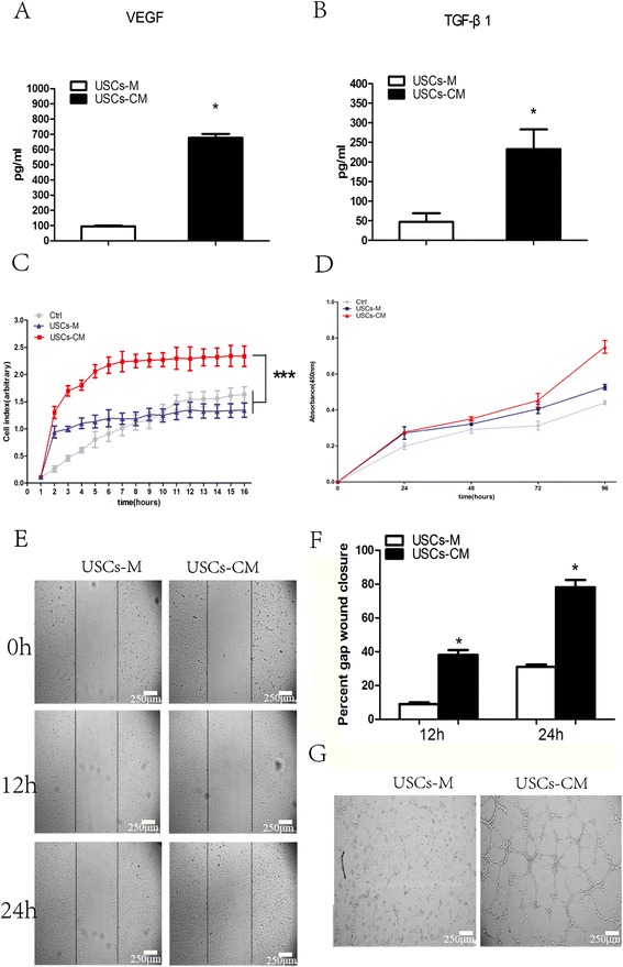
The ELISA assay for USCs-CM and the effect of USCs-CM on HUVECs. The concentration of VEGF (A) and TGF-β (B) in USCs-M and USCs-CM was measured by ELISA. (C) Real-Time Cell Analyzer showed USCs-CM significantly enhanced the migration of HUVECs. (D) CCK-8 assay showed USCs-CM significantly promoted HUVECs proliferation after 96 h incubation. (E and F) The scratch experiments demonstrated that USCs-CM increased the motility of HUVECs. (G) Large numbers of tube were formed in USCs-CM-cultured HUVECs, while no tube was formed in USCs-M-cultured HUVECs. *** P < 0.01 compared to the control or USCs-M groups at 16 h; *P < 0.05 compared to USCs-M groups. Scale bar = 250 μm.