
Figure 3.
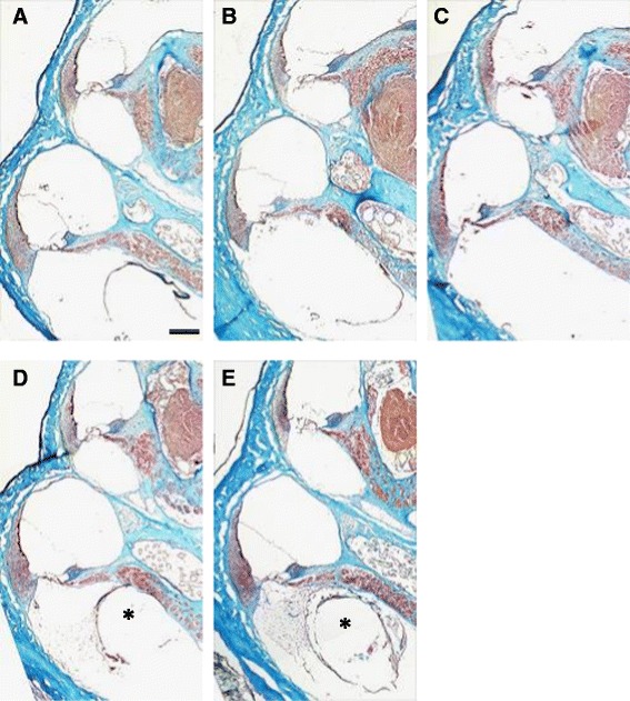
Representative photomicrographs of the guinea pig specimens. Mid-modiolar cochlear sections from groups A (non-coated), B (gelatin hydrogel coating), C (hydrogel + IGF1), D (hydrogel + HGF) and E (hydrogel + IGF1 + HGF). Third and basal turns of the cochlea are shown. In some cases, dense fibrous tissue was observed around the inserted electrode arrays (asterisks). Mallory’s trichrome staining. Scale bar, 250 μm.