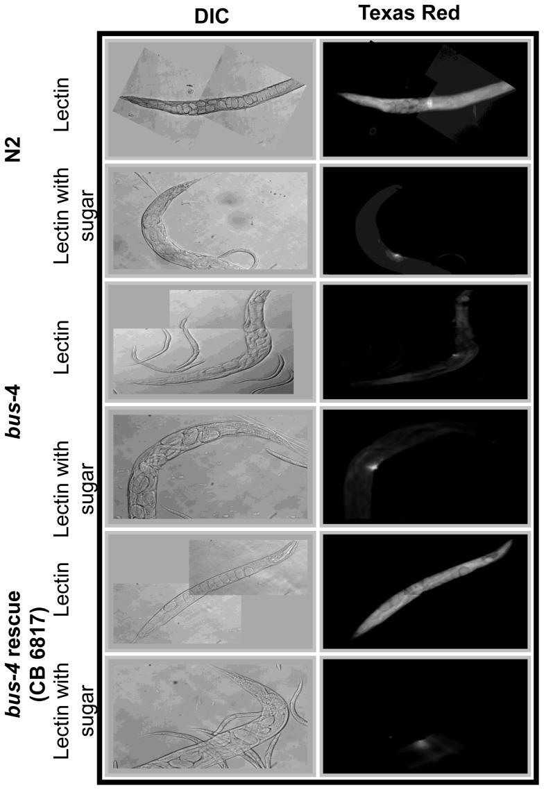
Figure 1. Lectin staining patterns of fixed bus-4 nematodes are altered.
Texas Red-conjugated Agaricus bisporus (ABA) Gal (β1,3) GalNAc specific lectin staining of whole mounted delipidated and live C. elegans strains are shown. A: the left columns shows differential interference contrast (DIC) micrographs and right columns show fluorescence micrographs of ABA stained fixed nematodes with and without prior incubation with inhibitory sugar (β-D-galactose). Top panels are N2 Bristol. Bottom panels are bus-4. B; Shorter exposures of ABA lectin are shown along with DIC micrographs to the left. Cuticle staining seen in the ventral tail region leading up to the anus in the N2 parent is absent in the bus-4 strain. C; Live ABA stained nematodes are shown. The surface coat of the bus-4 nematodes stains more intensely than in the N2 parent. The staining is most concentrated in the head and tail regions. Staining in the intestine is also increased abundances of soluble mucin-like proteins are indicated.

