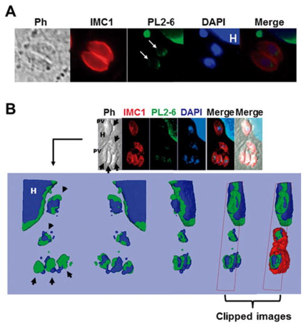
Fig. 3.
Immunolocalization of the epichromatin epitope in intracellular Toxoplasma gondii tachyzoites. (A) The image shows host cells infected with one parasitophorous vacuole (PV) containing two tachyzoites, sagittal plane. The epichromatin was identified by murine mAb antibody (PL2-6) stained with Alexa Fluor 488 (green). The panel (white arrows) shows an example representative of the typical PL2-6 labelling. IMC1, inner membrane complex 1 protein stained with Alexa Fluor 594 (red). DAPI was used to stain the nuclear DNA. PH, phase contrast; H, host cell; (B) Confocal section of an infected cell with three PV immunostained with mAb PL2-6, anti-IMC1 and DAPI staining. The arrows show single tachyzoites. The upper panel shows one slice of the Z projection, while the lower panel shows a 3D reconstruction (isosurface projection of the Z direction) of the entire stack in frontal, caudal and lateral view of the parasite. Colour coding: blue, the host (H) and parasite nuclei; green, the epichromatin. The last two representations on the right represent clipped images to demonstrate that epichromatin surrounds the parasite and host nuclei and that IMC surrounds epichromatin.

