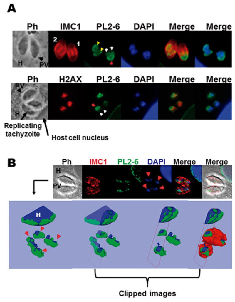
Fig. 5.

Immunolocalization of the epichromatin epitope in intracellular replicative tachyzoites. (A) The upper panel shows host cells infected with one parasitophorous vacuole (PV) containing two replicating tachyzoites in different degrees; numbers 1 and 2 indicate the lower and higher stage of replication, respectively. IMC1, inner membrane complex 1 protein stained with Alexa Fluor 594; PL2-6 epichromatin stained with Alexa Fluor 488 and DAPI used to dye the nuclear DNA. PH, phase contrast; H, host cell. The lower panel shows host cells infected with one PV containing three tachyzoites, one of them replicating. H2AX, variant T. gondii histone H2AX stained with Alexa Fluor 594; PL2-6, epichromatin stained with Alexa Fluor 488 and DAPI used to dye the nuclear DNA. Red arrow: the nuclear face facing the plasma membrane. Yellow head arrows: PL2-6 showing localization similar to that of mature tachyzoites in higher stage of replication. White head arrows: tops of the two nuclei entering the daughter cell; (B) One optical confocal section of an infected cell with one PV immunostained with mAb PL2-6 (Alexa Fluor 488); anti-IMC1 (Alexa Fluor 594) and DAPI staining. The arrows show the replicating nuclei of tachyzoites. The upper panel shows a 3D reconstruction (isosurface projection of the Z direction) of the entire stack of T. gondii culture. Colour coding: blue, the host (H) and parasite nuclei; green, the epichromatin. The last three representations on the right represent clipped images to demonstrate that epichromatin surrounds the parasite and host nuclei and that IMC surrounds epichromatin.
