Figure 2.
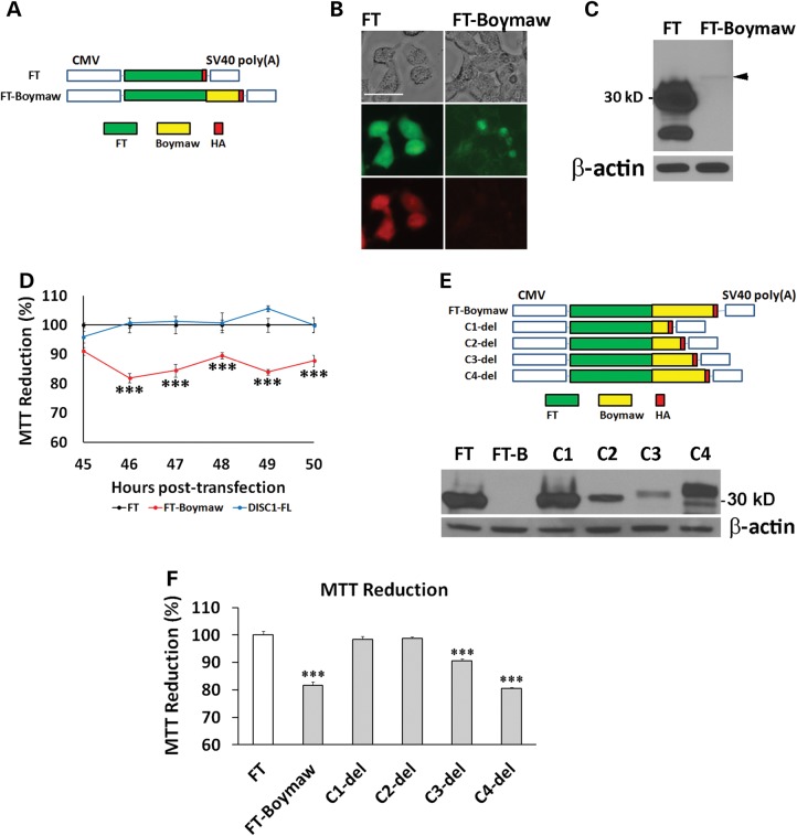
Characterization of the Boymaw gene. (A) Boymaw was fused with a FT tagged with HA in pTimer plasmid vector. The pTimer-HA plasmids (FT) were used as controls. (B) Expression of the FT-Boymaw fusion proteins was examined 48 h after transfection of HEK293T cells. While green and red fluorescence of the control FT proteins were very strong, little green and no red fluorescence of FT-Boymaw was detected. Scale bar: 15 µm. (C) Western blot analysis barely detected expression of the FT-Boymaw fusion proteins (black arrowhead) in comparison with abundant control FT proteins. (D) MTT assays revealed decreased MTT reduction by expression of the FT-Boymaw fusion gene in comparison with the controls and DISC1-FL [F(2,66) = 145.12, P < 0.0001]. Post hoc analyses (Tukey studentized range test) revealed that cells transfected with the FT-Boymaw construct displayed a significant decrease in MTT reduction at all time points except 45 h post-transfection. There were 4–6 replica wells per construct per time point. The mean value of the MTT reduction in cells transfected with FT was used as the control (100%) to calculate relative MTT reduction for the other two constructs at each time point. (E) A series of C-terminal deletion of FT-Boymaw fusion gene. Western blot analysis revealed expression of each deletion construct. (F) MTT assays were conducted 48 h after transfection of each deletion construct. There was a significant group effect on MTT reduction [F(8, 27) = 92.14, P < 0.000001]. Post hoc analyses (Tukey studentized range test) revealed significant MTT reduction in cells transfected with FT-Boymaw, C3-del and C4-del. There were four replica wells per construct. The mean value of the MTT reduction in cells transfected with FT was used as the control (100%) to calculate relative MTT reduction for all other constructs. Error bar: SEM. (*P < 0.05, **P < 0.01, ***P < 0.001).

