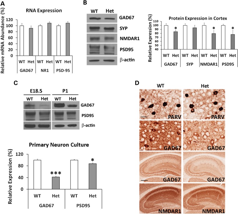
Figure 7.
Reduction of Gad67, Nmdar1 and Psd95 Proteins. (A) Q-PCR analysis of mRNA transcripts of Gad67, Nmdar1 and Psd95 genes in adult mouse brain. There was no significant difference between wild-type and the heterozygous DISC1-Boymaw mice in Gad67 [unpaired, two-tailed student's t-test, t(19) = 0.73, n.s.], Nmdar1 [unpaired, two-tailed student's t-test, t(19) = −1.44, n.s.] and Psd95 [unpaired, two-tailed student's t-test, t(19) = −1.17, n.s.] expression. (B) Western blot analysis and quantification of Gad67, synaptophysin (Syp), Nmdar1 and Psd95 in the brains of the adult heterozygous DISC1-Boymaw (n = 9) and wild-type (n = 9) mice. Unpaired two-tailed student's t-test was used for statistical analysis. No significant reduction of synaptophysin was observed between the two genotypes. Significant reduction of Gad67 [t(16) = 2.37, P < 0.05], Nmdar1 [t(16) = 2.55, P < 0.05] and Psd95 [t(16) = 2.58, P < 0.05] proteins was observed after normalization with either total amount of loading protein or β-actin expression. (C) In primary neurons isolated from cortex and striatum of postnatal Day 1 mice, significant reduction of both Gad67 (t(4) = 11.92, P < 0.001) and Psd95 [t(4) = 3.30, P < 0.05) proteins was observed in cultured primary neurons at 4 days in vitro from the DISC1-Boymaw mice (n = 3) in comparison with wild-type controls (n = 3) (unpaired, two-tailed student's t-test). Reduction of both proteins was further confirmed in the primary neuron culture (isolated from cortex and striatum) at 4 days in vitro from E18.5 embryos. (D) Immunohistochemical analysis of parvalbumin, Gad67, and Nmdar1 proteins in the heterozygous DISC1-Boymaw mice (anti-Psd95 antibodies did not work on paraffin sections). Reduction of Gad67 and Nmdar1 was observed in both hippocampus (scale bar: 100 µm) and cortex (scale bar: 20 µm). No difference was detected in parvalbumin expression. White ‘spots’ were nuclei not stained by antibodies. Error bar: SEM. (*P < 0.05, **P < 0.01, ***P < 0.001).

