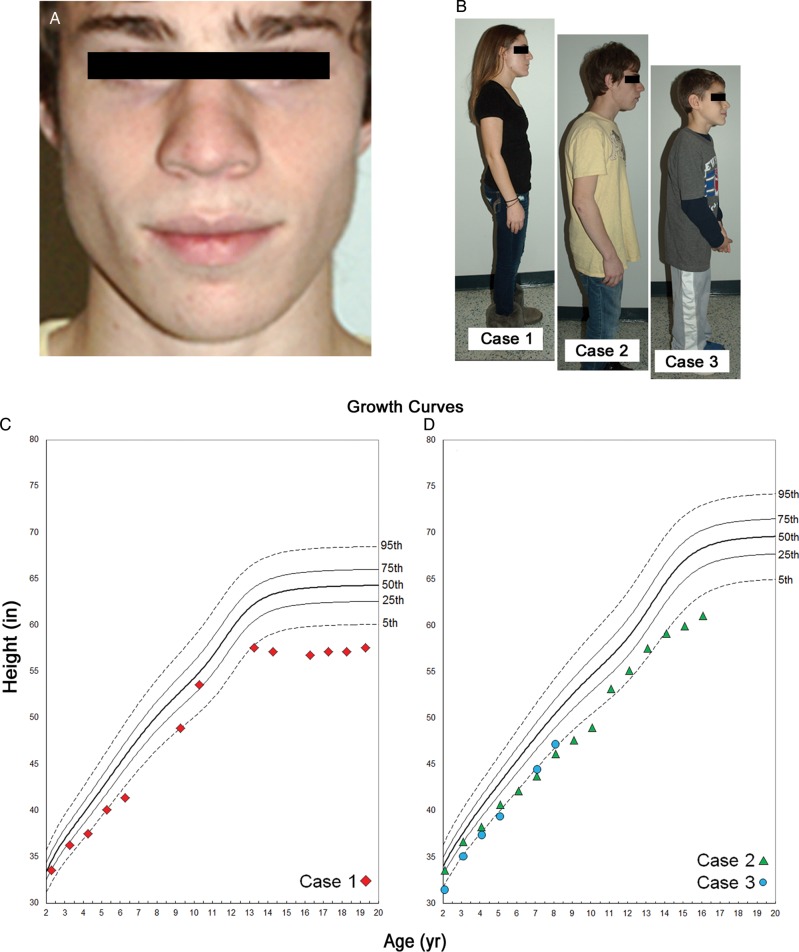
Figure 2.
Clinical presentation of the syndromic features in affected family members. Affected siblings shared distinct craniofacial and physical dysmorphologies. (A) Case 2, middle sibling, presented with prominent alae nasae and malar hypoplasia; the oldest and youngest sibling also exhibited these features in a less pronounced manner. (B) The body frame of each sibling suggests irregular physical development according to their respective ages. Growth curves (C, Case 1 and D, Cases 2 and 3) documenting stature from age 2 to the present illustrate a consistent pattern of abnormal height, falling below the 5th percentile of age- and gender-matched reference population.

