
Figure 3. Metacentrics have stronger centromeres relative to telocentrics in mice that have accumulated multiple metacentrics.
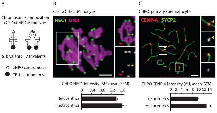
(A and B) Chromosome composition and HEC1 staining in CF-1 x CHPO MI oocytes (n=28). Staining was quantified at centromeres from CHPO telocentrics (identified as the dimmer kinetochores in bivalents, red asterisk) and from CHPO metacentrics in trivalents (yellow asterisk). (C) CENP-A staining in CHPO primary spermatocytes (n=67) was quantified for metacentrics (inset 1) and telocentrics (inset 2). Black asterisks: P<0.05; scale bars: 5 μm; AU: arbitrary units.