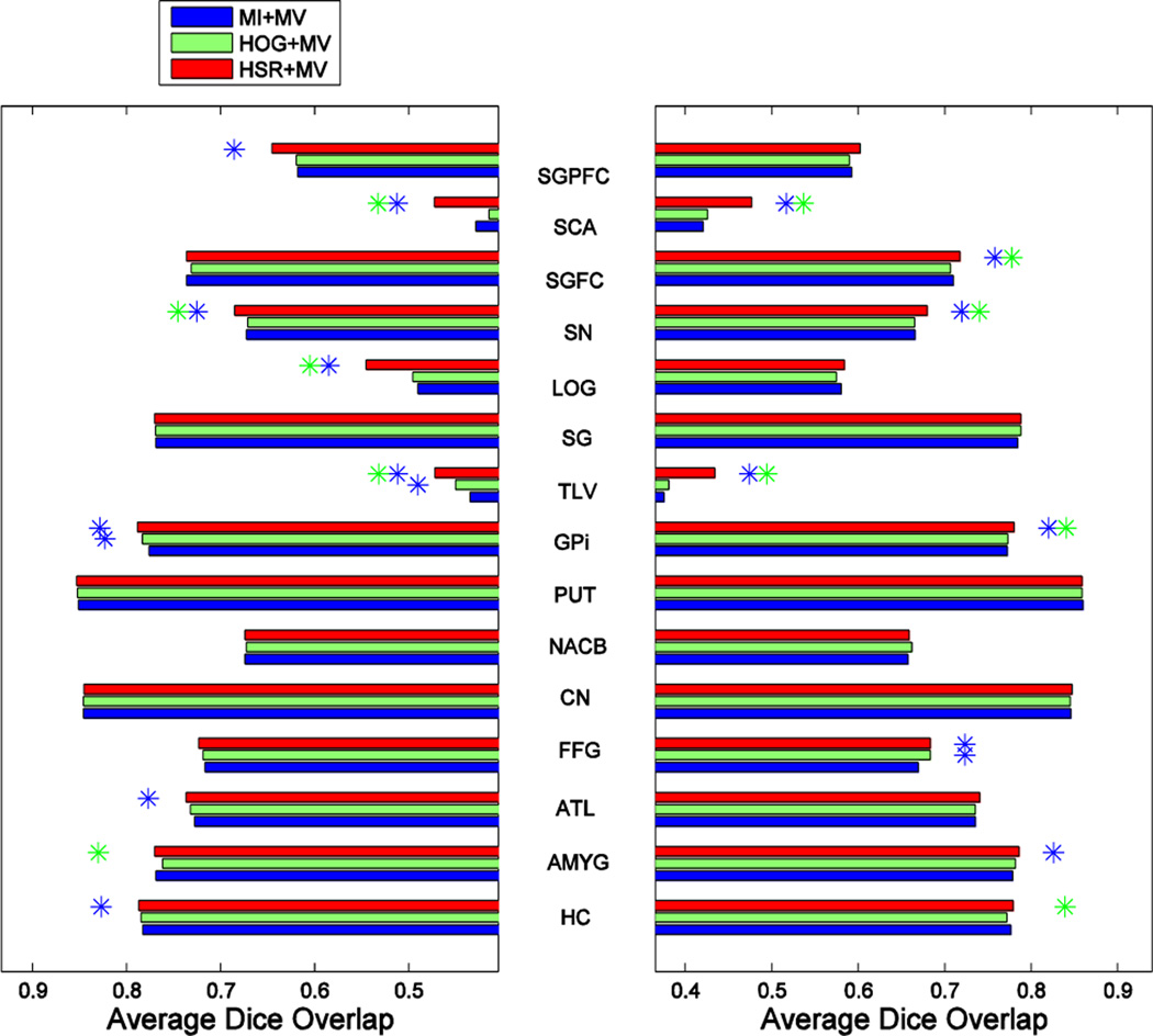
Fig. 12.
Blue, green and red bars show the segmentation performance, assessed by the Dice ratio, obtained by combining MV-based label fusion with different atlas selection methods on the labeling of 30 structures in the IXI dataset. Left and right plots show the results for the left and right parts of each structure, respectively, with the structure names shown in the middle. A blue and green asterisk represent significant improvement with respect to MI- and HOG-based selection, respectively.

