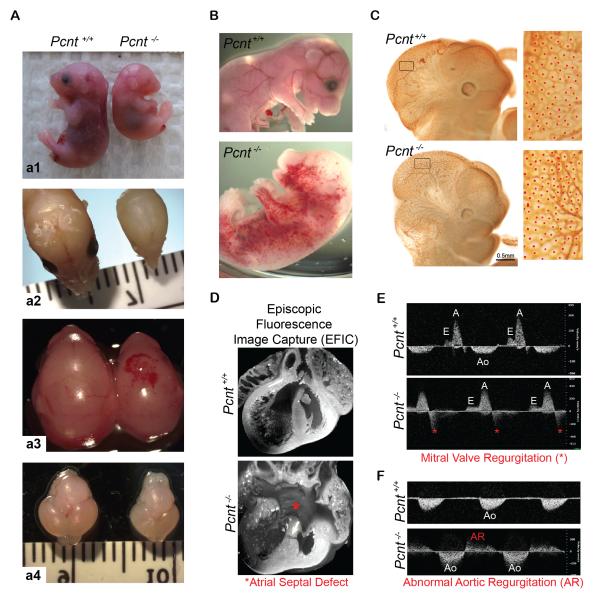
Figure 1. Pcnt−/− mice exhibit growth retardation, microcephaly, vascular anomalies and structural heart defects.
(A) Pcnt−/− mice exhibit intrauterine growth retardation (a1, 35% smaller, p<0.01; >50 mice in 7 liters); defects in cranial development (a2), intracranial hemorrhaging (a3); and microcephaly (a4).
(B) Representative images showing whole-body hemorrhaging in Pcnt−/− mouse (bottom) compared to the wild-type littermate (top).
(C) PECAM stain of vascular network in head of E11.5 Pcnt+/+ (top row) and Pcnt−/− (bottom row) embryos. Boxed regions (left column) are enlarged on the right, showing increased density of vascular plexuses (red dots) in Pcnt−/− head versus region-matched Pcnt+/+ (p<0.0001).
(D) 3-dimensional episcopic fluorescence image capture (EFIC) obtained from heart of E17.5 Pcnt+/+ (top) and Pcnt−/− (bottom) mice reveal defective septum formation (asterisk) in Pcnt−/− neonates in contrast to the Pcnt+/+ littermate.
(E-F) Doppler echocardiography reveals structural heart defects and congestive heart failure in Pcnt−/− embryos (bottom panels), but not in Pcnt+/+ littermates (top panels), as indicated by abnormal mitral valve regurgitation (asterisks, E) and aortic regurgitation (AR, F) at E15.5 and E17.5, respectively. Aortic flow (Ao); A wave, ventricular filling during atrial contraction (A); E wave, passive ventricular filling (E).

