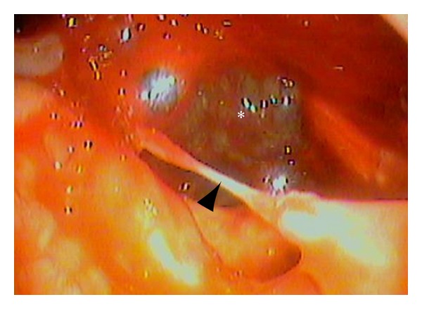
Figure 2.

A fiberscopic image showing the bleeding ulcer in the left lateral to the posterior part of the oropharynx. Note that the left of the figure indicates the medial side, lower anterior, since this is a view through an otolaryngological pharyngolaryngeal fiberscope. An ulcer is found from the left lateral wall of the oropharynx to the hypopharynx (asterisk). A clot is observed within the ulcer. The remnant mucosa in the ulcerated area formed a bridge-like structure (arrow heads).
