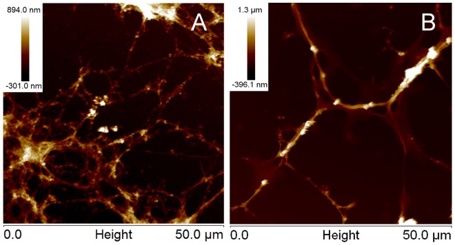
Figure 4. Overall morphology of polyLM and LM under AFM.
Atomic force microscopy images of polyLM (A) and LM (B) are shown in height mode after critical-point drying of the samples. Both matrices were obtained by incubating laminin with glass coverslips in the appropriate buffers for 12 hours. The scanned area was 2500 µm2.

