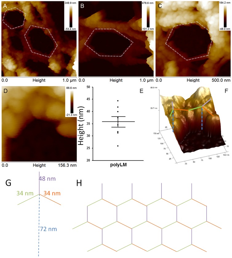
Figure 6. Atomic force microscopy reveals the occurrence of hexagonal-like figures in polyLM.
AFM was performed on polyLM matrices obtained as described in Figure 4 and areas of 1 (A, B) or 0.25 µm2 (C) were scanned in height mode. Hexagons-like figures similar to those occurring in natural laminin polymers [12] were identified. These hexagons were visible at different magnifications (A–C) and presented variable side lengths (sketched with white dashed lines), but they were never as short as 30 nm as they should be to correspond to the short arm of a laminin molecule. The smallest distinguishable structures contained within the sides of the hexagons were little globules (D) whose size and spacing was measured in images of 0.02 µm2 (D). Panel E shows the distribution of spacing values, which are compatible with the characteristic length (∼30 nm) of laminin polymerized via the short arms. Panel F depicts a three-dimensional reconstruction of the same area shown in panel D, with superposition of compatible locations of laminin molecules. Schemes of one individual laminin molecule (long arm dashed and short arms colored blue, green and orange), with indication of its characteristic dimensions (G) and of the hexagonal polymer generated by the interaction between individual laminin molecules (H) are also shown.

