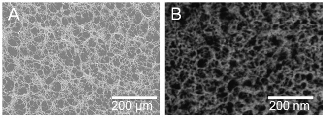
Figure 7. PolyLM displays similar morphologies at both 200 and 200,000 fold magnifications.

Images of polyLM were obtained using SEM (A) or transmission electron microscopy (B) after negative staining. Under SEM the magnification was 200 fold while under TEM it was 200,000 fold. Note that the observed patterns were alike despite the 1000 fold increase in magnification.
