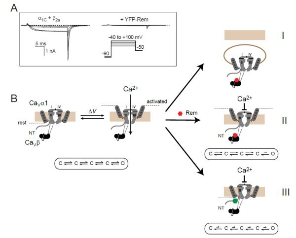
Figure 2. Rem inhibits CaV1.2 channels using multiple mechanisms.
(A) Left, exemplar whole-cell currents from HEK 293 cells expressing CaV1.2 α1C + β2a. Right, representative α1C + β2a channel currents in the presence of Rem. (B) Rem inhibits ICa using three independent mechanisms: by reducing channel surface density via enhanced dynamin-dependent endocytosis (mechanism I); by reducing channel Po independently of channel voltage sensor movement (mechanism II); by partially immobilizing channel voltage sensors as reported by a decrease in maximal gating charge (Qmax; mechanism III). Mechanisms I and II persist with RemT94N, suggesting they do not require GTP to be bound to the Rem nucleotide binding domain (represented as a red hexagon). By contrast, mechanism III is selectively lost with RemT94N suggesting a requirement for GTP binding to Rem (represented as a green hexagon). Figure modified from [57] and [17].

