Figure 1.
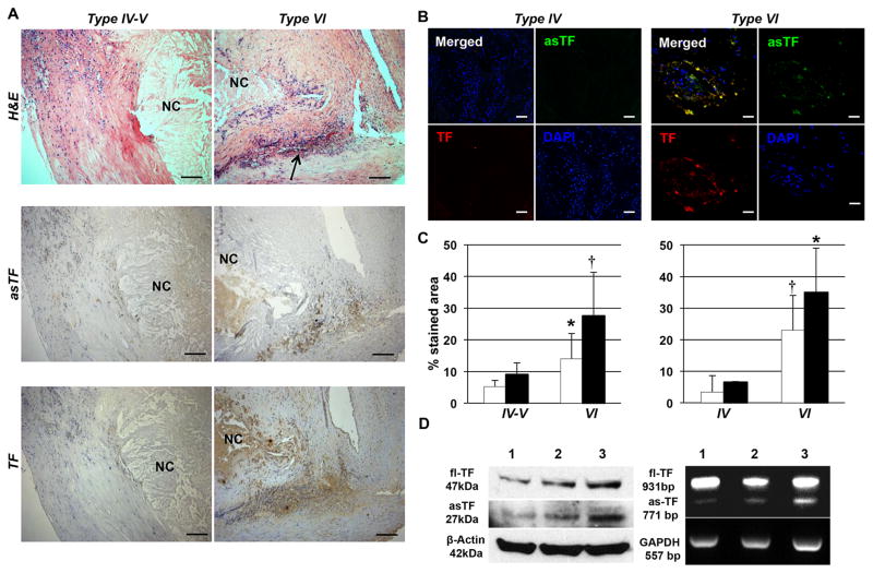
asTF expression is greater in type VI (complicated) lesions than type IV–V carotid and type IV coronary (uncomplicated) plaques. A. H&E stained sections (upper panel) of type IV–V (left; n=5) and type VI (right; n=5) carotid plaques. Necrotic Core (NC) and hemorrhage (arrow) of type VI lesions (right) are clearly visible. asTF (middle panel) and TF (bottom panel) immunostaining pattern of type VI (left) and IV–V (right) carotid plaques. Magnification: 100x. Scale bar=100 μm. B. Double immunofluorescence staining for asTF and TF of type IV (left; n=4) and VI (right; n=4) human coronary lesions. A greater expression of both asTF and TF and their co-localization was clear in Type VI vs. Type IV lesions. Magnification 400x. Scale Bar: 20 μm. C. Bars show asTF (white bars) and TF (black bars) quantification in type IV–V (n=5) and VI carotid (n=5; left panel) and in type IV (n=4) and VI coronary (n=4; right panel) plaques. *P=0.04 and †P=0.02 vs. type IV–V carotid or type IV coronary plaques. D. Western blot and PCR analysis of type IV–V (lines 1 and 2) and Type VI (line 3) carotid plaques shows asTF and fl-TF protein and mRNA expression in human plaques.

