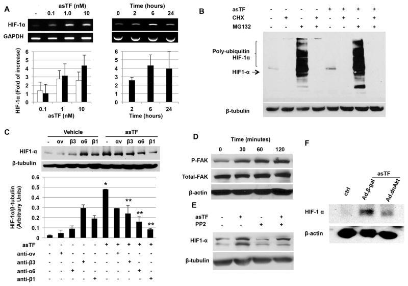
Figure 5.
Mechanisms involved in asTF-induced HIF-1α up-regulation. A. Dose-dependent up-regulation of HIF-1α mRNA by asTF was detected (left) at 2 hours (white bars) and 6 hours (black bars). Time course analysis of HIF-1α expression in the presence of asTF (10nM) shows a plateau at 6 hours (right). B. asTF enhanced protein synthesis of HIF-1α with no effect on degradation. C. Representative immunoblot (upper panel) shows inhibition of asTF-induced HIF-1α by integrin blockade. Bars (lower panel) show quantification of three separate experiments. *P<0.01 vs. vehicle. **P<0.01 vs. asTF. D. FAK activation by asTF was detected in endothelial cells stimulated with asTF. E. FAK inhibition by PP2 resulted in partial down-regulation of asTF-induced HIF-1α. F HIF-1α expression was inhibited in cells transduced with an adenovirus encoding dominant negative Akt (Ad.dnAkt) but not with Ad.βgal. Briefly, 24 hours following Ad.dnAkt or Ad.βgal (100 MOI) human aortic endothelial cells were treated with asTF (10nM) for 8 hours and HIF-1α expression measured by western blot. Data are the average of triplicates from single experiments that were independently repeated 3 times.

