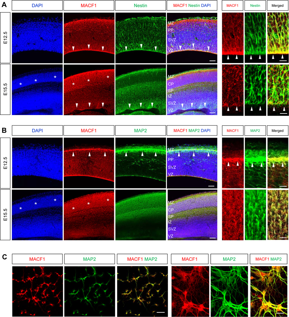
Figure 1. Expression of MACF1 in the developing cortex.
(A) Top panels: Immunostaining for MACF1 showed that the protein was broadly expressed in the developing mouse brain at E12.5. Notably, MACF1 was accumulated in the VZ where nestin-positive radial glial neural progenitors were present (arrow heads). Scale bar, 25 µm. Right three panels are higher magnification images showing co-localization of MACF1 and nestin. Scale bar, 10 µm. Bottom panels: At E15.5 stage, higher expression of MACF1 was found in the CP (stars), while the expression was reduced in radial glial progenitors at the VZ and the SVZ (arrow heads). Scale bar, 50 µm. Right three panels are higher magnification images. (B) Top panels: MACF1 protein was present in MAP2-positive cortical neurons in the MZ at E12.5 (arrow heads). Scale bar, 25 µm. Bottom panels: MACF1 was strongly expressed in the neurons of the CP and the SP at E15.5 (stars). Scale bar, 50 µm. Right three columns represent higher magnification images of upper cortex containing the MZ and PP (top), and the CP (bottom). Scale bar, 10 µm. MZ: marginal zone. PP: pre-plate. CP: cortical plate. SP: sub-plate. IZ: intermediate zone. SVZ: subventricular zone. VZ: ventricular zone. (C) MACF1 is expressed in neurites and somas of cortical neurons. Cortical neurons from E14.5 mice were cultured and immunostained with a MAP2 antibody. Scale bar, 50 µm. Right three panels are higher magnification images showing co-localization of MACF1 and MAP2. Scale bar, 25 µm.

