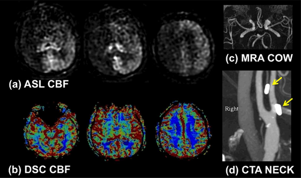
Figure 3.
(a) Example of poor PCASL labeling within the right anterior circulation due to poor labeling of the right internal carotid artery (ICA). Note the loss of ASL signal confined to this territory without compensatory collateral flow. In this case, confirmation was obtained with a (b) normal dynamic susceptibility contrast CBF map and (c) normal MR angiogram of the circle of Willis. (d) CT angiogram demonstrates surgical clips in the region of the right ICA (arrows), which may have been responsible for the poor labeling due to susceptibility effects. 60×35mm (300 × 300 DPI)

