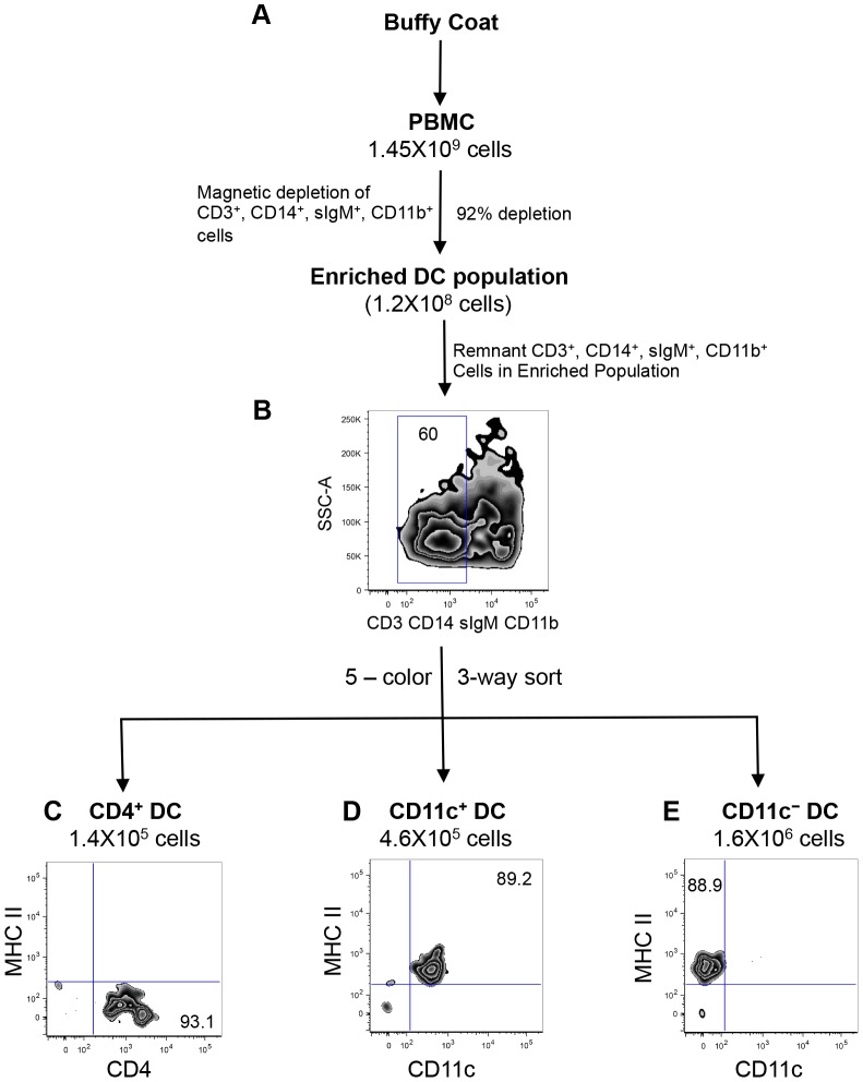
Figure 2. FACS purification of peripheral blood DC subsets.
Schematic diagram of the DC isolation protocol from PBMC. Following density centrifugation, PBMC were subjected to immuno-magnetic depletion of lineage positive cells, to enrich DC (A). To exclude remnant lineage positive cells present in the enriched DC population, a 5-color sort was performed using a BD FACS Aria II, according to the gating strategy shown in Figure 1A – F. Three major DC subsets that are MHC class II−/CD4+ (C) MHC class II+/CD11c+ (D), and MHC class II+/CD11c− (E). Numbers on plots represent percentage of cells. Data are representative of four independent experiments.

