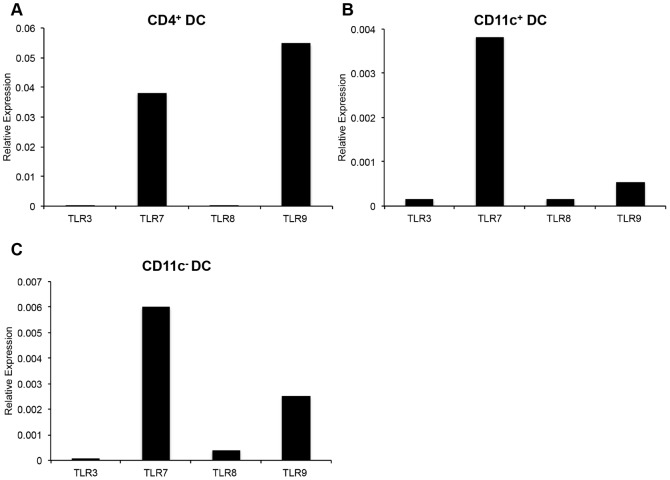
Figure 7. Real-time PCR quantification of TLR expression by peripheral blood DC subsets.
FACS purified CD4+ DC (A), CD11c+ DC (B), and CD11c− DC (C) were assessed for the expression of TLR3, TLR7, TLR8, and TLR9. Quantification of TLR expression was normalized to expression of GAPDH expression by DC subsets. Data are representative of two independent experiments.

