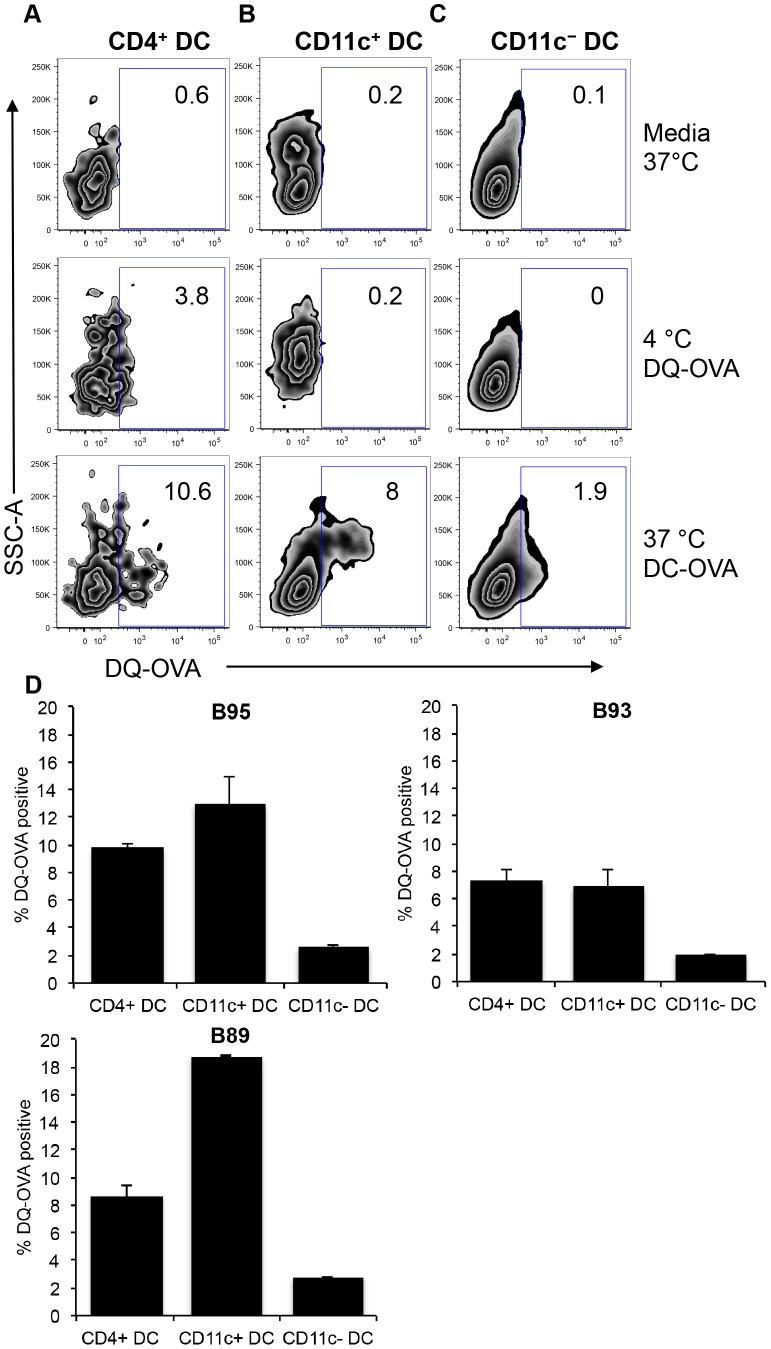
Figure 8. Internalization and degradation of exogenous antigen by peripheral blood DC subsets.
PBMC were incubated with self-quench fluorescent DQ-OVA for 1.5 hours at 4°C and 37°C. Cells were immuno-stained with surface antibodies to identify DC subsets as outlined in Figure 1. Dot plots show fluorescence of BODIPY that represents cleavage of DQ-OVA by CD4+ DC (A), CD11c+ DC (B), and CD11c− DC (C) from one animal. Calculation of DQ-OVA degradation efficiency by 3 different cattle was performed by subtracting BODIPY fluorescence of 4°C from 37°C (D). Error bars represent standard deviation.

