
Figure 7. LVS infection and miR-155 overexpression impair LPS-stimulated TNFα secretion.
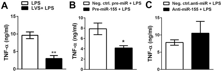
A. Control MDMs or cells that had been infected with LVS for 18 h were stimulated with 100 ng/ml E. coli LPS, and after 6 h at 37°C the amount of TNFα in the supernatant was quantified by ELISA. Data shown are the mean ±SEM (n = 3). B–C. MDMs were transfected with the indicated pre-miRs and anti-miRs 48 h prior to stimulation with E. coli LPS, and TNFα secretion was quantified as for panel A. Data are the mean ±SEM (n = 3).