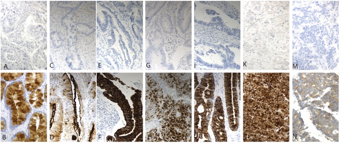
Figure 1. Immunohistochemical staining patterns of the antibiodies studied.
Representative images of antibody stainings in colorectal cancer; REG4-negative and -positive (A & B), MUC1- negative and -positive (C & D); MUC2-negative and -positive (E & F), MUC4-negative and -positive (G & H), MUC5AC-negative and -positive (I & J), synapthophysin-negative and -positive (K & L), chromogranin-negative and -positive (M & N) Original magnification was x 20.

