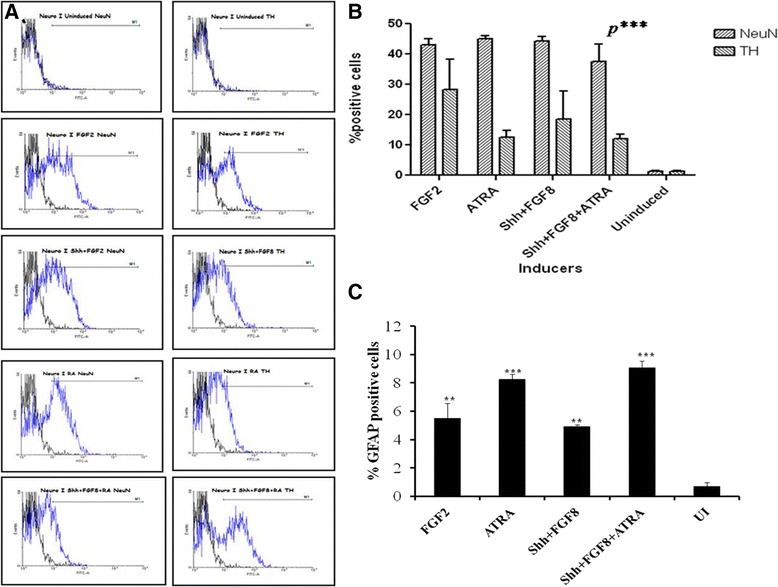
Figure 5.

Flow cytometric analysis of neuronal markers. A) Representative plots showing the expression of NeuN and TH in cells obtained from a single culture. B) Graph showing the% positive NeuN and TH cells in a single culture for all the induction media. C) Graph showing the% positive GFAP cells in all the induction media.
