
Figure 3.
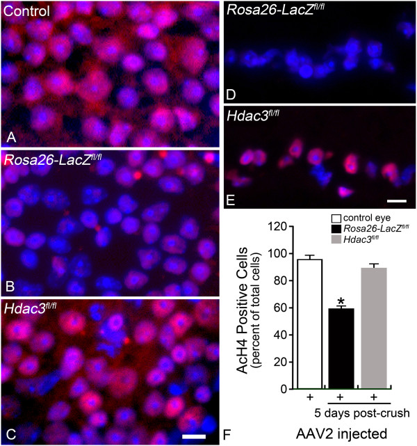
Hdac3 cKO ameliorated global deacetylation following RGC injury. (A-C) Retinal whole mounts were stained for AcH4 (red) in Rosa26-LacZ fl/fl and Hdac3 fl/fl AAV2-Cre/GFP injected and uninjected eyes at 5 days following optic nerve crush. The control retina was an uninjected and uncrushed Hdac3 fl/fl retina, which exhibited widespread AcH4 labeling (Scale bar: 10 μm). Nuclei absent of AcH4 staining were present only in the Rosa26-LacZ fl/fl retina after ONC. (D-E) Fluorescent microscopy of the retinal sections showed that the Hdac3 cKO GCL retained visibly more AcH4 labeled cells than the Rosa26-LacZ fl/fl GCL (Scale bar: 15 μm). (F) Cell counts in the GCL indicated Hdac3 cKO retinas at 5 days post ONC, retained AcH4 levels comparable to control retinas (P > 0.05), while Rosa26-LacZ fl/fl retinas exhibited about a 40% decrease in AcH4 labeled cells in the GCL compared to control retinas (*P ≤ 0.05).