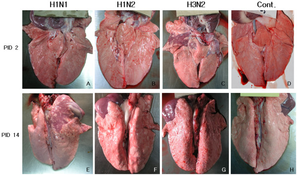
Figure 1.

Gross lung lesion of pigs infected with H1N1, H1N2, or H3N2 and non-infected pigs at post-inoculation days (PID) 2 and 14. The lesion typical for SIV presents as purple to dark red consolidated area in right cranial lobe of pig infected with H3N2 at PID 2 (C), however the lesion is not shown in the lung of the pig at PID 14 (G). The pig infected with H1N1 shows no lesion at PID 2 (A), but developed purple colored consolidated area in the right cranial lobe at PID 14 (E). The pigs infected with H2N1 shows had a small portion of purple consolidation in right caudal lobe at PID 2 (B), and mild broncho-pneumonia in both caudal lobes at PID 14 (F). Control pigs show no gross lesion at PID 2 (D) and at PID 14 (H).
