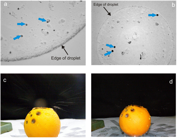
Figure 1.
(a). Conidia (blue bold arrows) in a 4 mm splashed droplet (edge indicated by fine arrow) observed under high power microscopy; (b). Three conidia (blue bold arrows) in a 1 mm droplet (edge indicated by fine arrow); (c). Splash emanating from infected orange (long exposure to show splash trajectories); (d). Splash droplets mid-flight (flash, short-term exposure).

