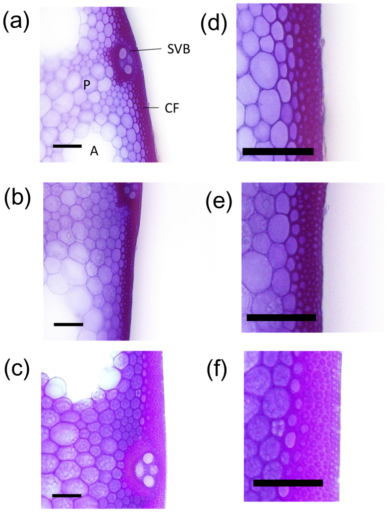
Figure 5. Morphological characteristics of transverse sections of cortical fiber tissues in culms.
(a, d) Koshihikari, (b, e) Chugoku 117, (c, f) Leaf Star. Scale bar: 100 μm. Transverse sections of fifth internodes 20 days after heading were stained with crystal violet. CF: cortical fiber tissue; SVB: small vascular bundle; P: parenchyma; A: aerenchyma.

