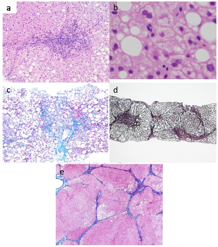
Figure 1.
Histological features of the different steps of fibrosis in NASH. (a) NASH: Steatosis, hepatocyte balooning degeneration, and inflammatory cells composed predominantly of lymphocytes in the portal area (Hematoxylin-eosin stain, 200× magnification); (b) Mallory body in NASH showed staghorn pattern (Hematoxylin-eosin stain, 400× magnification); (c) NASH: Detail of wire-mesh fibrosis and steatosis (Azan stain. 100× magnification); (d) Bridging fibrosis: Portal-central fibrous septa linking portal tracts and central veins (Reticulin silver stain, 40× magnification); (e) NASH-derived cirrhosis: Larger nodules with thin fibrous septa and steatosis (Azan stain. 40× magnification).

