Figure 2.

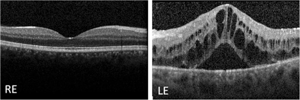
Baseline macular spectral domain optical coherence tomography. The examination reveals a normal macular morphology in the right eye and cystoid macular edema in the left eye. Abbreviations: LE, left eye; RE, right eye.

Baseline macular spectral domain optical coherence tomography. The examination reveals a normal macular morphology in the right eye and cystoid macular edema in the left eye. Abbreviations: LE, left eye; RE, right eye.