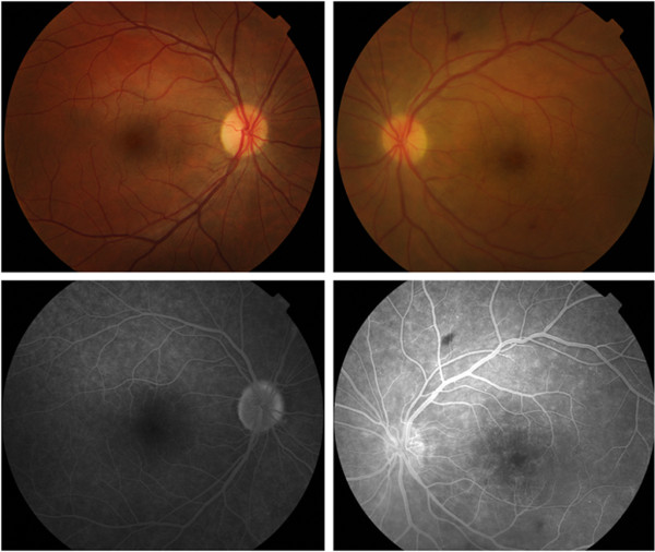
Figure 3.
Retinography and fundus fluorescein angiography following therapy with intravitreal bevacizumab. Right eye retinography (top left) reveals no pathologic alterations. Left eye retinography (top right) reveals optic disc and macular edema as well as a dot-blot hemorrhage near the inferotemporal arcade and a mid-peripheral flame-shaped hemorrhage. Right eye fluorescein angiography at 4'00" reveals staining of the optic disc margins (bottom left). Left eye fluorescein angiography at 3'30" reveals a reduction in macular and perivascular exudation in comparison to the previous examination; optic disc edema is also evident (bottom right).

