
Figure 7.
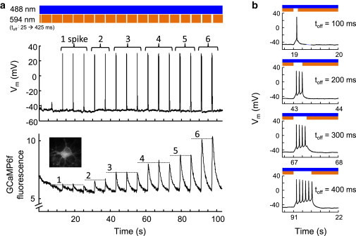
Crosstalk-free optical triggering of action potentials and optical monitoring of Ca2+ transients. (a) Calcium influx increased with the number of action potentials triggered by sdChR(C138S, E154A). (Top) Stoplight illumination scheme, with toff increasing from 25 to 425 ms. (Middle) Membrane voltage recorded in whole-cell current clamp. The number of action potentials induced in each off-period is labeled above the spike. (Bottom) Simultaneously recorded GCaMP6f fluorescence, showing spikes of discrete amplitude that grew with number of action potentials. (b) Closeups of the membrane voltage during the off-periods of increasing length. To see this figure in color, go online.