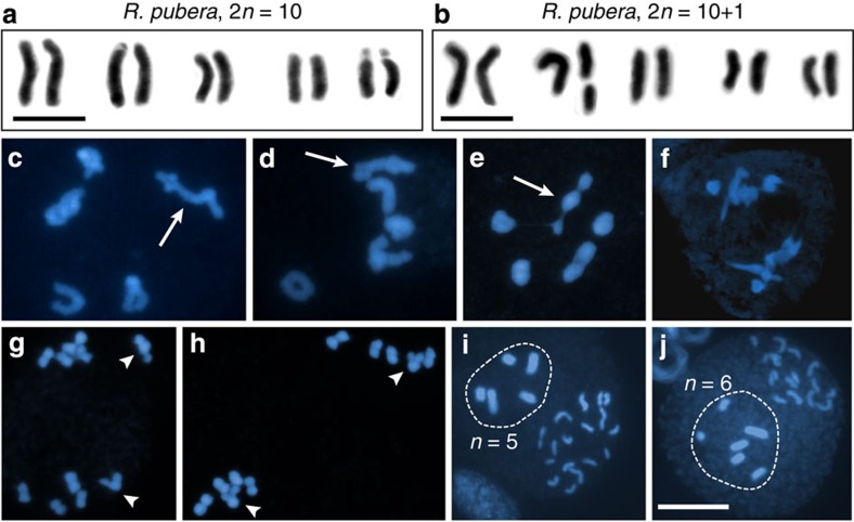
Figure 4. Meiosis of R. pubera (2n=10+1).
(a) Karyotype of R. pubera 2n=10. (b) Karyotype of R. pubera 2n=10+1. (c–j) DAPI images representing different stages of meiosis. (c,d) Cells in diakinesis showing the pairing behaviour of the heteromorphic bivalent (arrows). (e) Metaphase I. Arrow points to heteromorphic bivalent. (f) Anaphase I. (g,h) Cells in prophase II/metaphase II showing equational segregation of chromatids of the heteromorphic bivalent (arrowheads). (i,j) Microsporogenesis showing examples of pseudomonads in which the nucleus with (i) n=5 or (j) n=6 (highlighted) is centrally positioned and will give rise to a pollen grain. Size bars correspond to 10 μm.

