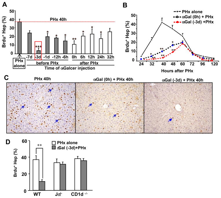
Fig. 1. Activation of iNKT cells by α-GalCer inhibits PHx-induced liver regeneration.
(A) Mice were treated with α-GalCer before or after PHx. All mice were euthanized 40h post PHx, and liver tissues were collected. BrdU was injected 2 h prior to euthanizing mice. The red rectangle indicates the greatest inhibition of hepatocyte proliferation. (B) Mice were treated with α-GalCer 3 d prior to PHx or concurrently (0 h) with PHx and euthanized at various time points post-PHx. BrdU was injected 2 h prior to euthanizing mice. (C) Representative BrdU immunohistochemical staining from panel B. (D) WT and NKT cell knockout mice were treated with or without α-GalCer for 3 d, and then PHx was performed. BrdU incorporation was determined 40 h post-PHx. The BrdU-positive hepatocytes in panels A, B, and D were counted in 5 high-power fields (100×), and the results are expressed as a percentage of total hepatocytes. The blue arrows in panel C indicate representative BrdU+ hepatocytes. Data represent the means ± SEM (n=6–10 mice). *P<0.05, **P<0.01, and ***P<0.001 compared with PHx alone.

